Synthetic community [SynCom] transfer for the...
Super-Resolution Microscopy of Functional Synaptic Vesicle Pools
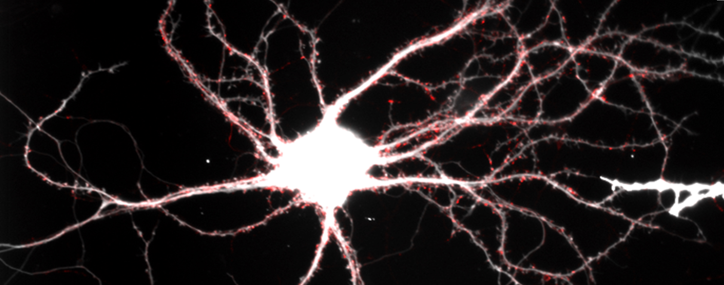
Neurotransmitter release from presynaptic vesicles at the synapse allows neurons to communicate in the brain. However the detailed spatial arrangement of different functional types of synaptic vesicles within the synapse remains unknown. We will use state-of-the-art super-resolution microscopy, including multi-colour, live and 3D STORM/STED microscopy, together with developing new analytical approaches, to address this question. We will then use these approaches to image changes occurring during synapse development, in response to neuronal activity and when key synaptic proteins are perturbed.
Disciplines and Techniques
Project supervisor/s
Dr. Laura Andreae
Laura is interested in neural stem cell manipulation, generation of brain organoids, structural and functional in vivo imaging, optogenetic manipulation of neural circuits,
King's College London
Dr. Susan Cox
Susan is interested in the development of new super-resolution localisation microscopy techniques, both through the development of optical systems and the creation of novel image analysis algorithms.
King's College University
References
Spontaneous Neurotransmitter Release Shapes Dendritic Arbors via Long-Range Activation of NMDA Receptors
Cell Rep, doi:10.1016/j.celrep .01.032
2015
The role of spontaneous neurotransmission in synapse and circuit development.
J Neurosci Res 96, 354-359, doi:10.1002/jnr.24154
2018
Independent vesicle pools underlie different modes of release during neuronal development
The Journal of neuroscience : the official journal of the Society for Neuroscience 32, 1867-1874, doi:10.1523/JNEUROSCI.5181-11.2012
2012
Bayesian localisation microscopy reveals nanoscale podosome dynamics
Nature Methods 9 195-200
2012
Super-resolution imaging in live cells
Dev Biol. 2015 May 1;401(1):175-81
2015