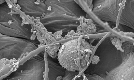


This is in part due to a lack of drug discovery platforms capable of assessing complex human neuromuscular disease phenotypes in a scalable manner. A major obstacle has been generating scaffolds to stabilise mature contractile myofibers in a multi-well assay format amenable to high content image analysis (HCI). This study describes the development of a scalable human iPSC-neuromuscular disease model, whereby suspended elastomer nanofibers support long-term stability, alignment, maturation, and repeated contractions of iPSC-myofibers, innervated by iPSC-motor neurons in 96-well assay plates. In this platform, optogenetic stimulation of the motor neurons elicits robust myofiber-contractions, providing a functional readout of neuromuscular transmission.
Additionally, HCI provides rapid and automated quantification of axonal outgrowth, myofiber morphology, and neuromuscular synapse number and morphology. By incorporating amyotrophic lateral sclerosis (ALS)-related TDP-43G298S mutant motor neurons and CRISPR-corrected controls, key neuromuscular disease phenotypes are recapitulated, including weaker myofiber contractions, reduced axonal outgrowth, and reduced number of neuromuscular synapses. Treatment with a candidate ALS drug, the receptor-interacting protein kinase-1 (RIPK1)-inhibitor necrostatin-1, rescues these phenotypes in a dose-dependent manner, highlighting the potential of this platform to screen novel treatments for neuromuscular diseases.
Neuromuscular diseases represent a diverse class of disorders with unmet clinical need. In several diseases such as amyotrophic lateral sclerosis (ALS) and spinal muscular atrophy (SMA) degeneration of motor neurons, the nerve cells that innervate skeletal muscle, leads to progressive paralysis and death[1-2]. Conversely, in muscular dystrophies, deterioration of neuromuscular function is caused by progressive weakness and wasting of the muscle[3] . Furthermore, in several autoimmune disorders, such as myasthenia gravis, degeneration of the neuromuscular junction (NMJ) itself, triggered by an autoimmune attack, leads to impaired movement[4] .
In many of these conditions peripheral axonal and synaptic dysfunction are key pathological events. Indeed, ALS is characterised by early degeneration of peripheral motor axons and neuromuscular synapses prior to cell death within the central nervous system (CNS) and progressive paralysis[1, 5] . Currently, there is no cure for this disease, yet growing evidence suggests that preserving neuromuscular synapses can extend lifespan in animal models and in patients[6-7] . 2D co-culture systems of primary motor neurons and myofibers which model nerve-muscle connectivity were first established in the 1970s[8], and later adapted to neuromuscular circuits containing mouse[9] and human[10] pluripotent stem cell-derived motor neurons. However, a major hurdle for applying such co-cultures to developing new treatments for axonal and synaptic degeneration in neuromuscular diseases is the lack of scalable in vitro disease models, amenable to high-throughput screening, that recapitulate complex disease phenotypes. A significant challenge has been stabilising mature contractile myofibers in a multi-well format that is suitable for automated high content image (HCI) analysis. In 2D cultures, contractile myofibers detach from the rigid tissue culture plate surface, precluding longitudinal phenotypic analysis[11-12] .
Several 3D solutions to this problem have involved suspending bundles of myofibers between flexible micropillars[13-17] or nylon hooks of VelcroTM fabric[18] , or attaching a hydrogel-embedded sheet of myofibers to flat polymer anchor points within compartmentalised tissue culture devices[19-21] , but the scalability of these approaches and amenability to automated HCI analysis has been limited. Nanofiber scaffolds have been used to successfully stabilise cardiomyocytes in multi-well assay plates[22] , however, in this instance, rigid attachment of the nanofibers to the tissue-culture surface precludes elastic recoil of the nanofibers upon muscle contraction, making this approach less suitable for culturing skeletal myofibers. Finally, while a number of approaches have focussed on generating scalable myogenic screening platforms in multi-well formats[16-17, 23], to our knowledge no neuromuscular co-culture platform has previously been generated in a 96-well assay format compatible with existing HCI analysis platforms.